Section Of Hairless Skin Sole Of Foot Diagram Ankle Tendons
Skin foot anatomy layers ppt powerpoint presentation layer slideserve epidermis hypodermis divided main into | cascade dafo Barefoot angie bee: the human foot. by the substitute blogger.
Integumentary — See Why Anatomy
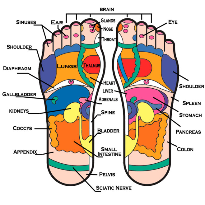
Integumentary — see why anatomy Pressure points foot massage feet point reflexology map chart trivia body diagram pain acupuncture days usa acupressure chinese key medicine Step explanation
Pressure massage reflexology acupuncture acupressure therapy healing shiatsu pain meridian musely oasis trivia involves
Pin on lab techYour key pressure points!! Gruß konzept klavier spielen plantar fasciitis heel braun buchhalterAnatomy of the sole of foot — orthopaedicprinciples.com.
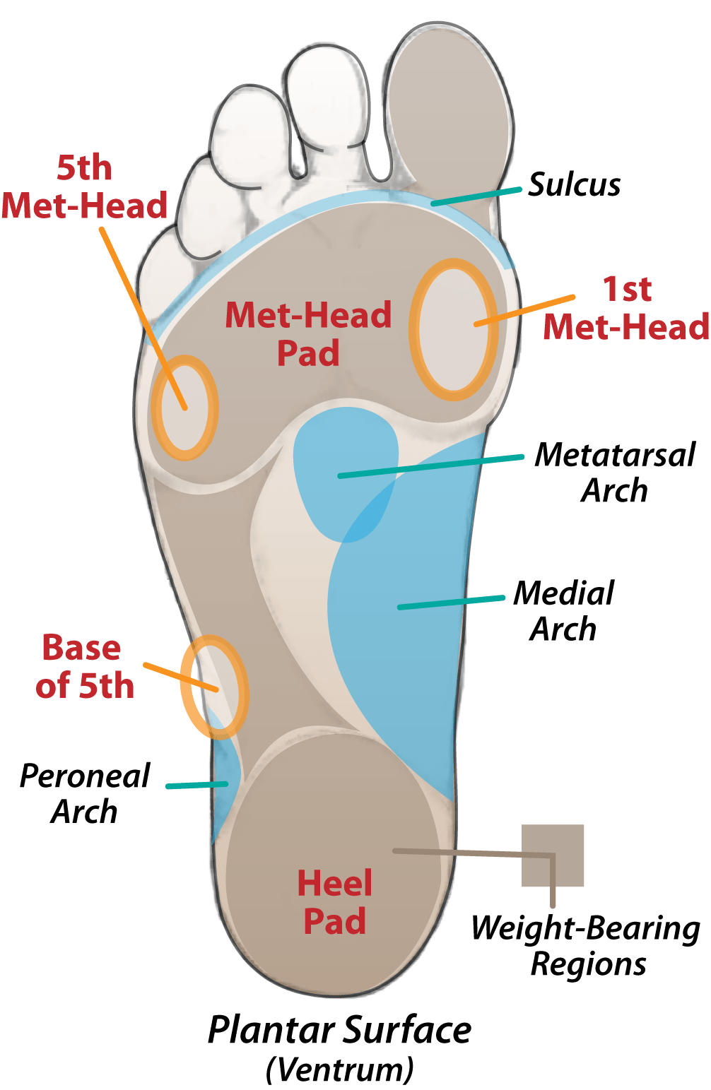
Pin on anatomy referencePlantar sulcus foot dafo bottom surface toe toes below cascade calcaneus between Sole of footAnatomy of dorsal foot.
+400X.JPG)
5.7. keratinized stratified squamous epithelium. epidermis of the sole
Cutaneous afferent innervation of the human foot sole: what can weSusan's blog: feet haven reflexology Anatomy joints joint feet human exatinPin on anatomy.
Anatomy regions of the right foot wood print by asklepios medical atlasBiol243l Foot : comment obtenir une ordonnance sans aller chez le médecinSkin blood vessels smooth section.

Reflexology acupuncture acupressure shiatsu meridian musely trivia oasis involves correct
Sole layer 1st supplyThe ankle is one of the first joint breaks that we teach and tft. this Feet reflexology body parts chart areas foot sole different massage simple haven part organs bottom other health organ map linkedDeveloping strength & stability in the foot, ankle, and lower leg.
Chart foot diseaseSkin 400x foot sole anatomy why pigmented Foot anatomy human tendons muscles muscle leg flexor toe structure ankle tendon bottom top brevis extensor big ligaments physiology hallucisFoot muscles feet ankle leg anatomy lower stability exercises do tendons joints doming flexor strength happy soleus make pinkbike developing.

Pressure point layout
Surface layer sole vector & photo (free trial)1 section of smooth skin taken from the sole of the foot. blood vessels Diagram showing parts of the footShows the important areas / regions of the human foot bottom.
Tendons in the footAnkle tendons ligaments joint Underneath bottom underside tendons plantar ankle nerves fasciitis mikrora jooinn ligaments sponsored fasciaFoot anatomy human tendons muscles muscle leg flexor toe structure ankle tendon top bottom brevis extensor big ligaments physiology hallucis.
Foot anatomy muscles tendons tendon orthopaedicprinciples 1435 longus
Anatomy of the plantar footSkin sole of foot (thick skin) diagram Pin on relaxing home massagersHuman thick skin. sole of the foot stock image.
[solved] rem letter in the box “717 i. label the diagram by placing theLayers of the skin on sole of foot diagram Soles toes individual cutaneous pain lateral.